Protecting families from contaminated food and water
All fields are required
Protecting families from contaminated food and water
Posted in Food Safety,Our Blog,Outbreaks & Recalls on October 10, 2024
After a year long investigation it has been found that undercooked bear meat leads to sickness. We often hear about people becoming ill from undercooked meats but we assume that it is the normal grocery store varieties of beef, pork, chicken etc. We don’t often let our minds wander to the consumption of wild game or animals harvested by hunting. The same rules apply, but sometimes even more so because there is generally no real inspection of the wild game meats before consuming.
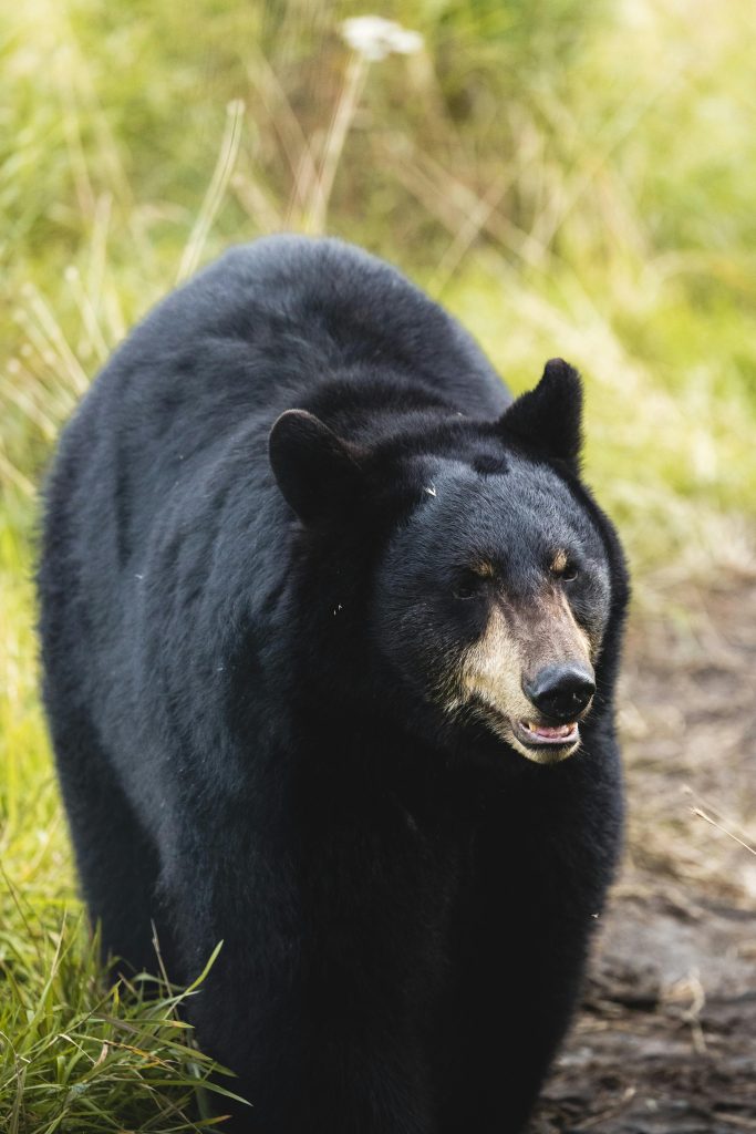
In a recent report it was found that the cause of 10 people becoming ill after an event in North Carolina was in fact undercooked bear. The CDC identified the likely sickness as trichinellosis, a parasitic infection that causes muscle pain, fever and facial swelling. It comes from eating undercooked animals containing trichinella larvae. One of the most common hosts is the black bear, which is found in the western region of the state where this “gathering” took place. In this case, a majority of the people who got sick were under 18 years old, with six minors reporting illness in the weeks following the event.
Nine of the 10 people infected had facial swelling. Six had muscle pain and four had fevers, according to the CDC. “Because black bears are common hosts for Trichinella spp., communicating methods for properly cooking and preparing wild game meat is important,” the CDC said in a statement. Officials recommend cooking game meat to an internal temperature of at least 165°F (74°C) to kill parasites, emphasizing that “freezing might not be sufficient” to prevent infection. Though rare, trichinellosis can lead to death in 0.2% of cases.
Trichinellosis (trichinosis) is caused by nematodes (roundworms) of the genus Trichinella. In addition to the classical agent T. spiralis (found worldwide in many carnivorous and omnivorous animals), several other species of Trichinella are now recognized, including T. pseudospiralis (mammals and birds worldwide), T. nativa (Arctic bears), T. nelsoni (African predators and scavengers), T. britovi (carnivores of Europe and western Asia), and T. papuae (wild and domestic pigs, Papua New Guinea and Thailand). Trichinella zimbabwensis is found in crocodiles in Africa but to date there are no known associations of this species with human disease.
Depending on the classification used, there are several species of Trichinella: T. spiralis, T. pseudospiralis, T. nativa, T. murelli, T. nelsoni, T. britovi, T. papuae, and T. zimbabwensis, all but the last of which have been implicated in human disease. Adult worms and encysted larvae develop within a single vertebrate host, and an infected animal serves as a definitive host and potential intermediate host. A second host is required to perpetuate the life cycle of Trichinella. The domestic cycle most often involved pigs and anthropophilic rodents, but other domestic animals such as horses can be involved. In the sylvatic cycle, the range of infected animals is great, but animals most often associated as sources of human infection are bear, moose and wild boar.
Trichinellosis is caused by the ingestion of undercooked meat containing encysted larvae (except for T. pseudospiralis and T. papuae, which do not encyst) of Trichinella species. After exposure to gastric acid and pepsin, the larvae are released from the cysts and invade the small bowel mucosa where they develop into adult worms. Females are 2.2 mm in length; males 1.2 mm. The life span in the small bowel is about four weeks. After 1 week, the females release larvae that migrate to striated muscles where they encyst. Diagnosis is usually made based on clinical symptoms, and is confirmed by serology or identification of encysted or non-encysted larvae in biopsy or autopsy specimens.
Light infections may be asymptomatic. Intestinal invasion can be accompanied by gastrointestinal symptoms (diarrhea, abdominal pain, vomiting). Larval migration into muscle tissues (one week after infection) can cause periorbital and facial edema, conjunctivitis, fever, myalgias, splinter hemorrhages, rashes, and peripheral eosinophilia. Occasional life-threatening manifestations include myocarditis, central nervous system involvement, and pneumonitis. Larval encystment in the muscles causes myalgia and weakness, followed by subsidence of symptoms.
For more information on outbreak information and food safety tips please follow Make Food Safe.